SCI Publications
2015
E. Erdil, A.O. Argunsah, T. Tasdizen, D. Unay, M. Cetin.
“A joint classification and segmentation approach for dendritic spine segmentation in 2-photon microscopy images,” In 2015 IEEE 12th International Symposium on Biomedical Imaging (ISBI), IEEE, April, 2015.
DOI: 10.1109/isbi.2015.7163992
Shape priors have been successfully used in challenging biomedical imaging problems. However when the shape distribution involves multiple shape classes, leading to a multimodal shape density, effective use of shape priors in segmentation becomes more challenging. In such scenarios, knowing the class of the shape can aid the segmentation process, which is of course unknown a priori. In this paper, we propose a joint classification and segmentation approach for dendritic spine segmentation which infers the class of the spine during segmentation and adapts the remaining segmentation process accordingly. We evaluate our proposed approach on 2-photon microscopy images containing dendritic spines and compare its performance quantitatively to an existing approach based on nonparametric shape priors. Both visual and quantitative results demonstrate the effectiveness of our approach in dendritic spine segmentation.
T. Etiene, R.M. Kirby, C. Silva.
“An Introduction to Verification of Visualization Techniques,” Morgan & Claypool Publishers, 2015.
SCI Institute.
Note: FluoRender: An interactive rendering tool for confocal microscopy data visualization. Scientific Computing and Imaging Institute (SCI) Download from: http://www.fluorender.org, 2015.
Note: FusionView: Problem Solving Environment for MHD Visualization. Scientific Computing and Imaging Institute (SCI), Download from: http://www.scirun.org, 2015.
Y. Gao, L. Zhu, J. Cates, R. S. MacLeod, S. Bouix,, A. Tannenbaum.
“A Kalman Filtering Perspective for Multiatlas Segmentation,” In SIAM J. Imaging Sciences, Vol. 8, No. 2, pp. 1007-1029. 2015.
DOI: 10.1137/130933423
M.U. Ghani, S.D. Kanik, A.O. Argunsah, T. Tasdizen, D. Unay, M. Cetin.
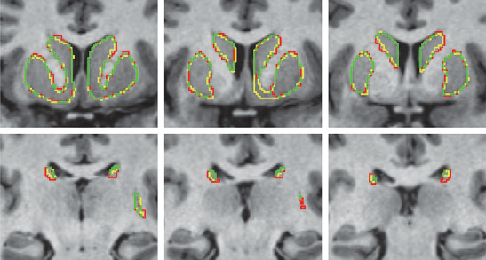
“Dendritic spine shape classification from two-photon microscopy images,” In 2015 23nd Signal Processing and Communications Applications Conference (SIU), IEEE, May, 2015.
DOI: 10.1109/siu.2015.7129985
Functional properties of a neuron are coupled with its morphology, particularly the morphology of dendritic spines. Spine volume has been used as the primary morphological parameter in order the characterize the structure and function coupling. However, this reductionist approach neglects the rich shape repertoire of dendritic spines. First step to incorporate spine shape information into functional coupling is classifying main spine shapes that were proposed in the literature. Due to the lack of reliable and fully automatic tools to analyze the morphology of the spines, such analysis is often performed manually, which is a laborious and time intensive task and prone to subjectivity. In this paper we present an automated approach to extract features using basic image processing techniques, and classify spines into mushroom or stubby by applying machine learning algorithms. Out of 50 manually segmented mushroom and stubby spines, Support Vector Machine was able to classify 98% of the spines correctly.
K. Gillette, J.D. Tate, B. Kindall, P. Van Dam, E. Kholmovski, R.S. MacLeod.
“Generation of Combined-Modality Tetrahedral Meshes,” In Computing in Cardiology, 2015.
Registering and combining anatomical components from different image modalities, like MRI and CT that have different tissue contrast, could result in patient-specific models that more closely represent underlying anatomical structures.
In this study, we combined a pair of CT and MRI scans of a pig thorax to make a tetrahedral mesh and compared different registration techniques including rigid, affine, thin plate spline morphing (TPSM), and iterative closest point (ICP), to superimpose the segmented bones from the CT scan on the soft tissues segmented from the MRI. The TPSM and affine-registered bones remained close to, but not overlapping, important soft tissue.
Simulation models, including an ECG forward model and a defibrillation model, were computed on generated multi-modality meshes after TPSM and affine registration and compared to those based on the original torso mesh.
B. D. Goodwin, C. R. Butson.
“Subject-Specific Multiscale Modeling to Investigate Effects of Transcranial Magnetic Stimulation,” In Neuromodulation: Technology at the Neural Interface, Vol. 18, No. 8, Wiley-Blackwell, pp. 694--704. May, 2015.
DOI: 10.1111/ner.12296
OBJECTIVE:
Transcranial magnetic stimulation (TMS) is an effective intervention in noninvasive neuromodulation used to treat a number of neurophysiological disorders. Predicting the spatial extent to which neural tissue is affected by TMS remains a challenge. The goal of this study was to develop a computational model to predict specific locations of neural tissue that are activated during TMS. Using this approach, we assessed the effects of changing TMS coil orientation and waveform.
MATERIALS AND METHODS:
We integrated novel techniques to develop a subject-specific computational model, which contains three main components: 1) a figure-8 coil (Magstim, Magstim Company Limited, Carmarthenshire, UK); 2) an electromagnetic, time-dependent, nonhomogeneous, finite element model of the whole head; and 3) an adaptation of a previously published pyramidal cell neuron model. We then used our modeling approach to quantify the spatial extent of affected neural tissue for changes in TMS coil rotation and waveform.
RESULTS:
We found that our model shows more detailed predictions than previously published models, which underestimate the spatial extent of neural activation. Our results suggest that fortuitous sites of neural activation occur for all tested coil orientations. Additionally, our model predictions show that excitability of individual neural elements changes with a coil rotation of ±15°.
CONCLUSIONS:
Our results indicate that the extent of neuromodulation is more widespread than previous published models suggest. Additionally, both specific locations in cortex and the extent of stimulation in cortex depend on coil orientation to within ±15° at a minimum. Lastly, through computational means, we are able to provide insight into the effects of TMS at a cellular level, which is currently unachievable by imaging modalities.
C. Gritton, M. Berzins, R. M. Kirby.
“Improving Accuracy In Particle Methods Using Null Spaces and Filters,” In Proceedings of the IV International Conference on Particle-Based Methods - Fundamentals and Applications, Barcelona, Spain, Edited by E. Onate and M. Bischoff and D.R.J. Owen and P. Wriggers and T. Zohdi, CIMNE, pp. 202-213. September, 2015.
ISBN: 978-84-944244-7-2
While particle-in-cell type methods, such as MPM, have been very successful in providing solutions to many challenging problems there are some important issues that remain to be resolved with regard to their analysis. One such challenge relates to the difference in dimensionality between the particles and the grid points to which they are mapped. There exists a non-trivial null space of the linear operator that maps particles values onto nodal values. In other words, there are non-zero particle values values that when mapped to the nodes are zero there. Given positive mapping weights such null space values are oscillatory in nature. The null space may be viewed as a more general form of the ringing instability identified by Brackbill for PIC methods. It will be shown that it is possible to remove these null-space values from the solution and so to improve the accuracy of PIC methods, using a matrix SVD approach. The expense of doing this is prohibitive for real problems and so a local method is developed for doing this.
A. V. P. Grosset, M. Prasad, C. Christensen, A. Knoll, C. Hansen.
“TOD-Tree: Task-Overlapped Direct send Tree Image Compositing for Hybrid MPI Parallelism,” In Eurographics Symposium on Parallel Graphics and Visualization (2015), Edited by C. Dachsbacher, P. Navrátil, 2015.
A. Gunduz, H. Morita, P. J. Rossi, W. L. Allen, R. L. Alterman, H. Bronte-Stewart, C. R. Butson, D. Charles, S. Deckers, C. de Hemptinne, M. DeLong, D. Dougherty, J. Ellrich, K. D. Foote, J. Giordano, W. Goodman, B. D. Greenberg, D. Greene, R. Gross, J. W. Judy, E. Karst, A. Kent, B. Kopell, A. Lang, A. Lozano, C. Lungu, K. E. Lyons, A. Machado, H. Martens, C. McIntyre, H. Min, J. Neimat, J. Ostrem, S. Pannu, F. Ponce, N. Pouratian, D. Reymers, L. Schrock, S. Sheth, L. Shih, S. Stanslaski, G. K. Steinke, P. Stypulkowski, A. I. Tröster, L. Verhagen, H. Walker, M. S. Okun.
“Proceedings of the Second Annual Deep Brain Stimulation Think Tank: What's in the Pipeline,” In International Journal of Neuroscience, Vol. 125, No. 7, Taylor & Francis, pp. 475-485. 2015.
DOI: 10.3109/00207454.2014.999268
PubMed ID: 25526555
The proceedings of the 2nd Annual Deep Brain Stimulation Think Tank summarize the most contemporary clinical, electrophysiological, and computational work on DBS for the treatment of neurological and neuropsychiatric disease and represent the insights of a unique multidisciplinary ensemble of expert neurologists, neurosurgeons, neuropsychologists, psychiatrists, scientists, engineers and members of industry. Presentations and discussions covered a broad range of topics, including advocacy for DBS, improving clinical outcomes, innovations in computational models of DBS, understanding of the neurophysiology of Parkinson's disease (PD) and Tourette syndrome (TS) and evolving sensor and device technologies.
A. Gyulassy, A. Knoll, K. C. Lau, Bei Wang, P. T. Bremer, M. E. Papka, L. A. Curtiss, V. Pascucci.
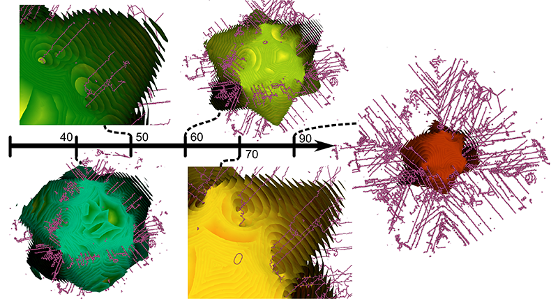
“Morse-Smale Analysis of Ion Diffusion for DFT Battery Materials Simulations,” In Topology-Based Methods in Visualization (TopoInVis), 2015.
Ab initio molecular dynamics (AIMD) simulations are increasingly useful in modeling, optimizing and synthesizing materials in energy sciences. In solving Schrodinger's equation, they generate the electronic structure of the simulated atoms as a scalar field. However, methods for analyzing these volume data are not yet common in molecular visualization. The Morse-Smale complex is a proven, versatile tool for topological analysis of scalar fields. In this paper, we apply the discrete Morse-Smale complex to analysis of first-principles battery materials simulations. We consider a carbon nanosphere structure used in battery materials research, and employ Morse-Smale decomposition to determine the possible lithium ion diffusion paths within that structure. Our approach is novel in that it uses the wavefunction itself as opposed distance fields, and that we analyze the 1-skeleton of the Morse-Smale complex to reconstruct our diffusion paths. Furthermore, it is the first application where specific motifs in the graph structure of the complete 1-skeleton define features, namely carbon rings with specific valence. We compare our analysis of DFT data with that of a distance field approximation, and discuss implications on larger classical molecular dynamics simulations.
A. Gyulassy, A. Knoll, K. C. Lau, Bei Wang, PT. Bremer, M.l E. Papka, L. A. Curtiss, V. Pascucci.
“Interstitial and Interlayer Ion Diffusion Geometry Extraction in Graphitic Nanosphere Battery Materials,” In Proceedings IEEE Visualization Conference, 2015.
J. K. Holmen, A. Humphrey, M. Berzins.
“Exploring Use of the Reserved Core,” In High Performance Parallelism Pearls, Edited by J. Reinders and J. Jeffers, Elsevier, pp. 229-242. 2015.
DOI: 10.1016/b978-0-12-803819-2.00010-0
In this chapter, we illustrate benefits of thinking in terms of thread management techniques when using a centralized scheduler model along with interoperability of MPI and PThreads. This is facilitated through an exploration of thread placement strategies for an algorithm modeling radiative heat transfer with special attention to the 61st core. This algorithm plays a key role within the Uintah Computational Framework (UCF) and current efforts taking place at the University of Utah to model next-generation, large-scale clean coal boilers. In such simulations, this algorithm models the dominant form of heat transfer and consumes a large portion of compute time. Exemplified by a real-world example, this chapter presents our early efforts in porting a key portion of a scalability-centric codebase to the Intel ® Xeon PhiTM coprocessor. Specifically, this chapter presents results from our experiments profiling the native execution of a reverse Monte-Carlo ray tracing-based radiation model on a single coprocessor. These results demonstrate that our fastest run confiurations utilized the 61st core and that performance was not profoundly impacted when explicitly over-subscribing the coprocessor operating system thread. Additionally, this chapter presents a portion of radiation model source code, a MIC-centric UCF cross-compilation example, and less conventional thread management techniques for developers utilizing the PThreads threading model.
A. Humphrey, T. Harman, M. Berzins, P. Smith.
“A Scalable Algorithm for Radiative Heat Transfer Using Reverse Monte Carlo Ray Tracing,” In High Performance Computing, Lecture Notes in Computer Science, Vol. 9137, Edited by Kunkel, Julian M. and Ludwig, Thomas, Springer International Publishing, pp. 212-230. 2015.
ISBN: 978-3-319-20118-4
DOI: 10.1007/978-3-319-20119-1_16
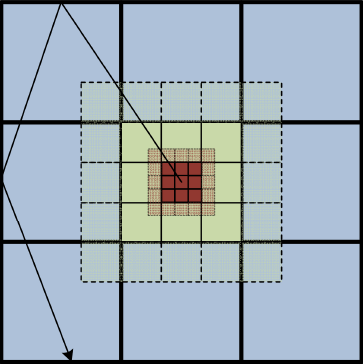
Keywords: Uintah; Radiation modeling; Parallel; Scalability; Adaptive mesh refinement; Simulation science; Titan
CIBC.
Note: ImageVis3D: An interactive visualization software system for large-scale volume data. Scientific Computing and Imaging Institute (SCI), Download from: http://www.imagevis3d.org, 2015.
C.R. Johnson, K. Potter.
“Visualization,” In The Princeton Companion to Applied Mathematics, Edited by Nicholas J. Higham, Princeton University Press, pp. 843-846. September, 2015.
ISBN: 9780691150390
H. De Sterck, C.R. Johnson.
“Data Science: What Is It and How Is It Taught?,” In SIAM News, SIAM, July, 2015.
C.R. Johnson.
“Computational Methods and Software for Bioelectric Field Problems,” In Biomedical Engineering Handbook, 4, Vol. 1, Ch. 43, Edited by J.D. Bronzino and D.R. Peterson, CRC Press, pp. 1--28. 2015.
Computer modeling and simulation continue to become more important in the field of bioengineering. The reasons for this growing importance are manyfold. First, mathematical modeling has been shown to be a substantial tool for the investigation of complex biophysical phenomena. Second, since the level of complexity one can model parallels the existing hardware configurations, advances in computer architecture have made it feasible to apply the computational paradigm to complex biophysical systems. Hence, while biological complexity continues to outstrip the capabilities of even the largest computational systems, the computational methodology has taken hold in bioengineering and has been used successfully to suggest physiologically and clinically important scenarios and results.
This chapter provides an overview of numerical techniques that can be applied to a class of bioelectric field problems. Bioelectric field problems are found in a wide variety of biomedical applications, which range from single cells, to organs, up to models that incorporate partial to full human structures. We describe some general modeling techniques that will be applicable, in part, to all the aforementioned applications. We focus our study on a class of bioelectric volume conductor problems that arise in electrocardiography (ECG) and electroencephalography (EEG).
We begin by stating the mathematical formulation for a bioelectric volume conductor, continue by describing the model construction process, and follow with sections on numerical solutions and computational considerations. We continue with a section on error analysis coupled with a brief introduction to adaptive methods. We conclude with a section on software.
C.R. Johnson.
“Visualization,” In Encyclopedia of Applied and Computational Mathematics, Edited by Björn Engquist, Springer, pp. 1537-1546. 2015.
ISBN: 978-3-540-70528-4
DOI: 10.1007/978-3-540-70529-1_368
Page 35 of 144
