Even as cancer treatment options are multiplying and becoming more sophisticated, one relatively straightforward approach will always be in play: cutting the diseased tissue out of the body.
One persistent challenge with such cancer surgeries is preserving as much of the affected organ as possible while also ensuring all cancerous cells are removed. The so-called “margins” of a cancerous mass are hard to concretely define even when looking directly through a microscope, and are even more ambiguous in the kind of non-invasive scans patients undergo before surgery.
As such, surgeons must use their eyes and best judgment to know how much tissue to remove — all while the patient is under the knife. The consequences of error here are serious precisely because they are not immediately apparent: there is little way to tell that cancerous cells remain growing inside the patient until they begin forming new tumors.
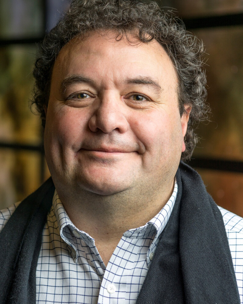
Valerio Pascucci , Professor in Price Engineering’s Kahlert School Of Computing, is now collaborating with researchers at Tulane University and the University of Georgia on a new Biden Cancer Moonshot project that aims to revolutionize this aspect of cancer treatment.
Today, President Joe Biden and First Lady Jill Biden are announcing an up to $23 million award to create an imaging system that will give doctors the ability to scan a tumor during surgery and determine within minutes whether any cancer tissue has been left behind. While patients are still under anesthesia in the operating room doctors will be able to assure patients, with certainty, that all the cancer has been removed from the area, making repeated invasive surgeries unnecessary.
The funding for the award the Bidens are announcing comes from the Advanced Research Projects Agency for Health (ARPA-H), a federal funding agency established by the Biden Administration to rapidly advance high-potential, high-impact biomedical research that cannot be readily accomplished through traditional research or commercial activity.
Tulane researchers will lead a team focused on overcoming the technical computing and engineering challenges to make the advanced imaging device a reality within the next five years. Receiving the full $23 million will require the team to reach certain milestones in their efforts.
“Currently, it can take days to weeks before a surgeon knows whether all the tumor has been removed, and our goal is to get that down to 10 minutes, while the patient is still on the table,” says J. Quincy Brown, associate professor of biomedical engineering in the Tulane School of Science and Engineering and lead researcher on the project. “If successful, our work would transform cancer surgery as we know it.”
The project, called MAGIC-SCAN (Machine-learning Assisted Gigantic Image Cancer margin SCANner), would be one of the world’s fastest high-resolution tissue scanners, capable of detecting residual cancer cells on the surface of removed organs within minutes. The system would be trained on thousands of clinical scans so that it can accurately highlight cancer at a cellular level as it renders a highly detailed 3D map of the surface of the tumor. Once the map is complete, surgeons could then match cancerous margins with their counterparts inside the surgical site, removing additional tissue as necessary.
Tulane researchers have already been working on developing this technology using prostate and colorectal cancer patients – two of the most difficult kinds of tumors to remove – and they’ve managed to get the detection time down to about 45 minutes.
Pascucci and his colleagues at the U will work on the cyber infrastructure required to handle the massive sets of data from patients needed to train the machine-learning models. Called FASTMAP for “Fast, Accelerated Support for Training MAchine Learning models on Petascale data,” this element of the project is emblematic of the work of the U’s Scientific Computing and Imaging Institute, where Pascucci is a faculty member.
“FASTMAP is a human-centered approach that innovates responsible AI for cancer research by adopting the National Science Data Fabric infrastructure in two key areas,” Pascucci said. “First, FASTMAP will introduce a high-performance computing cyberinfrastructure capable of repeatedly developing and training new AI models over petascale data in a matter of days for continuous feedback and validation by clinical pathologists.
“Second,” he said. “FASTMAP will develop highly precise AI models that can be executed within minutes in the operating room, providing real-time guidance to surgeons during surgery.”
Collaborating with Tulane and the U will be researchers from the University of Georgia, who will work on improving the quality of the imaging resolution.
Clinical validation of the device will be accomplished with partners at Cedars-Sinai Medical Center in Los Angeles, Southeast Louisiana Veterans Hospital and East Jefferson General Hospital. The Tulane-spinout company Instapath Inc. will help the team develop FDA-compliant versions of the new scanner. The project is part of a broader initiative by ARPA-H to develop Precision Surgical Interventions (PSI) that improve surgical accuracy and reduce errors.
The University of Utah is home to Huntsman Cancer Institute, a National Cancer Institute-designated Comprehensive Cancer Center. Huntsman Cancer Institute hosted then-Vice President Biden in 2016 as he kicked off the National Cancer Moonshot.
“MAGIC-SCAN represents the highest calling of engineers and lies at the core of the U’s mission,” says Charles Musgrave, Dean of the Price College of Engineering. “There’s no better way to serve the people of Utah, the nation, and the world than to turn raw data into action that saves lives.”
Original article from the John and Marcia Price College of Engineering

